![]()
| This study was done by a group of researchers interested in the effect of the reovirus on bladder cancer cells (published in 2003, see reference below). |
![]()
|
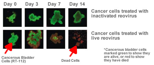
The first set of images shows what cells look like when they are treated in a special way to show green if they are still alive, or red if they have been killed. The first row of pictures shows cancer cells treated with inactivated reovirus (in effect, not treated and growing in a normal way that cancer grows). Notice the second row shows substantial cell death of cancerous cells when treated with the live reovirus compared to the first row showing cancerous cell growth.
|
![]()
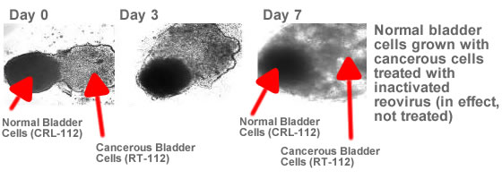
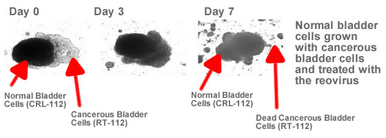
These researchers went much farther with this study and decided to test the theory that the reovirus would kill cancer cells but leave normal cells alone. The first set of images shows the normal growth of cancerous bladder cells grown together with non-cancerous bladder cells.
The second image shows the effect of treating the combination of normal and cancerous cells with the reovirus. Notice substantial cell killing rather then growth in a fairly short period of time. |
|
Finally, the researchers wanted to be sure that the reovirus was not having an effect on normal bladder cells. This last set of images shows that there is no discernable effect when normal bladder cells are treated with either live or killed reovirus.
|
![]()
|
Source: doi:10.1016/S0168-1702(03)00045-5 Selective reovirus killing of bladder cancer in a co-culture spheroid model Ruhangiz T. Kilani(a), Yahya Tamimi(a), Erich G. Hanel(a), Kevin K. Wong(a), Shahzeer Karmali(a), Patrick W. K. Lee(b) and Ronald B. Moore(a). (a) Divisions of Experimental Surgery and Oncology, Department of Surgery,
University of Alberta and Department of Oncology, Cross Cancer Institute,
Edmonton, AB, Canada T6G 1Z2 |