|
|
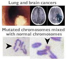
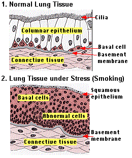
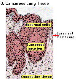
Consider lung cancer. The pictures on the left (click for source) represent the growth of lung cancer in a chronic smoker. The basal cells in normal lung tissue (Graphic 1 on left) represent the stem cells for the columnar epithelium, which is the interior lining of the lungs. This is the tissue most exposed to smoke in a cigarette. Due to cellular stress caused by the toxins in cigarette smoke, the basal cells naturally divide to provide a thicker layer of cells against the toxins (Graphic 2 - after approx. 10 years smoking). The top layer of cells flatten out and form a more protective barrier also. It is almost like the lining of the lungs, is becoming more like your skin to protect you from the toxins. The problem is that many of these basal cells are mutants with a very abnormal nucleus. In time, some mutations happen that cause the cells to produce protease proteins to melt through the basement membrane of the lung tissue (Graphic 3 - after approx. 20 years smoking) and start to invade other tissue. Most of these cells secrete a mucous similar to the basement membrane to protect the group from the immune system. Others will occassionally produce the protease that allow them to break from the group through the mucous and invade additional tissue. Reovirus represents a natural enemy of growing cancers. The lead cancerous cells, secret proteases on their growing edges that melt cell junctions and membranes in front of them. These same proteases melt the outer protective protein coat of the reovirus making it more likely to infect the cell. The cancer cells that produce the mucous also have a problem with Reovirus. An uncoated reovirus particle exposes a sigma1 attachment fiber that can drill through this mucous to reach and infect the cell. Once inside the cancer cell, the Reovirus may invoke cell death and signalling of the immune system through apoptosis, or, it may continue to divide and destroy the cell itself. The response in the cell depends alot on the type of cell mutations it contains. It is thought that a mutation in the Ras cell pathway is enough to signal apoptosis and the immune cells. Further, addtional mutations though Ras/RalGEF/p38 cell pathway determine if Reovirus can replicate (click for source). The normal cellular defense against reovirus (activated PKR) appears to be controlled by this pathway. Finally, the dead or dying cancer cells are likely to attract the immune system. Should these cells show cell surface features or other things that are abnormal, the immune system will be alerted to watch out for these cell types. |